Wednesday April 30, 2008
Board style question for Critical Care fellows.
Case: 42 yr old male admitted with Guillain-Barré Syndrome and intubated due to rapidly falling vital capacity. Patient otherwise remain fairly stable and sedated with average dose of 5 mg/kg/hr Propofol. Unfortunately, patient failed 5 days of Plasma exchange therapy. On day 6, pt develop exacerbation of his baseline asthma and was started on IV solumedrol but steroids were discontinued next day on neurology’s recommendation as it may prolong recovery from GBS. All labs and clinical exam otherwise remain stable including mental status which was assessed briefly each morning while off sedation. DVT and GI prophylaxis on place. Enteral feeding started on day 2. Bedside percutaneous trach and PEG has been planned.
While doing 'shift' on night of day 7, you noticed some downward BP "trend" but as labs and exam so far remain rock stable, you attributed it to sedation. While browsing 5 AM labs you noticed PH of 7.25 and bicarb of 14. Chem-7 showed Cr of 2.1 (baseline 1.1) and K of 5.7. As you get more attentive to patient, you noticed frequent episodes of bradycardia on monitor. Tracking back monitor in last few hours showed multiple alarms for bradycardia but was 'silenced' as this was the most stable patient in unit. Also pulse ox trend from upper 90s to lower 90s. You ordered lactate level, cardiac enzymes, EKG, CXR, broad spectrum antibiotics, panculture, adjust ventilator and gave IVF bolus. Lactate level is back with 7.2 and indeed patient has NSTE MI with Troponin-I of 7.1. CPK is reported in 5 K range. You called primary. Cardiology, Nephrology and ID services were put on consult. Pt. required another 2 IVF boluses before you left at 7 AM. Pt. continue to deteriorate and died 48 hours later despite combined endeavor of all services to salvage his hemodynamic collapse.
Your diagnosis (choose one).
A) Acute MI from plasma exchange therapy.
B) Acute septic shock due to use of steroid.
C) Side effect of propofol.
D) Acute renal failure from hypotension.
E) Ventilator associated pneumonia
Answer is C: Propofol infusion syndrome.
As propofol has gained enormous popularity in ICUs, it is extremely important to be aware of "Propofol infusion syndrome", particularly when drip is continued for more than 48 hours.
Syndrome consist of myocardial failure, metabolic acidosis, renal failure, lipemia, rhabdomyolysis, and hyperkalemia. Clues to "Propofol infusion sundrome" are unexplained lactate level, bradycardia and increasing need for pressors. It’s a clinical diagnosis.
Due to poorly understood reason, syndrome is associated with acute neurological illnesses or acute inflammatory diseases and receiving steroids in addition to propofol.
Also independent syndrome consist of bronchospasm, hypotension and anaphylactic type picture has been reported with start of infusion apart from "propofol infusion syndrome" above.
Some critics blame high lipid content of infusion for syndrome.
A) is wrong as acute MI is associated with IVIG theraphy for GBS and unlikely with plasma exchange. Also, this patient finished his therapy 2 days ago.
B) is wrong as there is no clear evidence of sepsis and short term use of steroid has less likely reason for acute sepsis. But please note that it is very important to practice aseptic technique while handling propofol.
D) is possible but extreme hypotension is unlikely to go unnoticed and doesn't explain all the clinical features.
E) VAP is not associated with this clinical picture
Wednesday, April 30, 2008
Tuesday, April 29, 2008
Tuesday April 29 , 2008
Carina as a Radiographic Landmark for Positioning the IABP
There are reports in literature that targeting tip of Intraaortic Balloon Pump (IABP) just below the aortic knob on CXR radiologically may still cause occlusion of left subclavian artery (upto 7 - 16%). 1,2.
See this interesting another approach to target carina as a Useful Radiographic Landmark for Positioning the Intraaortic Balloon Pump.
METHODS: The distance from the top of the distal aortic arch (aortic knob) to the left subclavian artery (LSCA) on three-dimensional computed tomography angiography in 100 patients, was measured. The distance from the level of the LSCA origin to the level of the carina was also measured using three-dimensional computed tomography in 150 additional patients.
RESULTS: In 16% of the aortic knob study population, the LSCA to aortic knob distance was <0 cm or 0 cm. The median distance from the LSCA to the carina was 42 mm (range: 30–63 mm). In the carina study population, the origin of the LSCA was 35–55 mm above the carina in 95.3% of patients.
CONCLUSION:
In 16% of patients, the IABP was too close to the LSCA origin when it was placed at the aortic knob, whereas Positioning the IABP at 2 cm above the carina provided an adequate position for the IABP tip (1.5–3.5 cm distal to the origin of the LSCA) in 95.3% of patients.
The carina may be a more reliable landmark for positioning the IABP than the aortic knob.
Reference: click to get abstract / article
1. AORTIC KNOB; CAN BE A RELIABLE RADIOGRAPHIC LANDMARK FOR PLACEMENT OF INTRA-AORTIC BALLOON PUMP TIP? - Canadian Journal of Anesthesia 53:26056 (2006)
2. The Carina as a Useful Radiographic Landmark for Positioning the Intraaortic Balloon Pump - Anesth Analg 2007;105:735-738
Monday, April 28, 2008
Monday April 28 , 2008
Prone Position: Is it still on in ARDS patients
Recently published meta- analysis of thirteen trials that enrolled a total of 1559 patients were studied. In 10 of those trials (n=1486) they found that prone positioning did not reduce mortality among hypoxemic patients. The lack of effect of ventilation in the prone position on mortality was similar in trials of prolonged prone positioning and in patients with acute lung injury.
Even though it does not reduce the mortality, but it did increase the oxygenation by 34% on day 1 in 8 of the trials. In 5 trials (n=1004), prone positioning was associated with a reduced risk of ventilator associated pneumonia.
Conclusion: Mechanical ventilation in the prone position does not reduce mortality or duration of ventilation despite improved oxygenation and a decreased risk of pneumonia.
Reference: click to get abstract / article
Sud S, Sud M, Friedrich JO, Adhikari NKJ. Effect of mechanical ventilation in the prone position on clinical outcomes in patients with acute hypoxemic respiratory failure: a systematic review and meta-analysis. CMAJ 2008; 178(9), full text here
Sunday, April 27, 2008
Sunday April 27 , 2008
Eosinopenia as a reliable marker of sepsis ?
See this interesting observation from a 12 bed medical ICU !
Introduction: The marked reduction in number of circulating eosinophil leucocytes in acute infection was first described by Zappert in 1893, and was utilized during the first quarter of the last century as a useful diagnostic sign.
Methods: Eosinophils were measured at ICU admission. Two intensivists blinded to eosinophils classified patients as
1. Eosinopenia is a reliable marker of sepsis on admission in medical intensive care units - Critical Care 2008, 12:R59, Full article in pdf available here
Eosinopenia as a reliable marker of sepsis ?
See this interesting observation from a 12 bed medical ICU !
Introduction: The marked reduction in number of circulating eosinophil leucocytes in acute infection was first described by Zappert in 1893, and was utilized during the first quarter of the last century as a useful diagnostic sign.
Methods: Eosinophils were measured at ICU admission. Two intensivists blinded to eosinophils classified patients as
- negative
- systemic inflammatory response syndrome (SIRS),
- sepsis,
- severe sepsis, and
- septic shock
Results: A total of 177 patients were enrolled.
A: In discriminating between non-infected (negative+SIRS) and infected (sepsis+severe sepsis+septic shock) groups,
Eosinophils less than 50cells/mm^3 yielded a
- sensitivity 80% and specificity 91%,
positive likelihood ratio (LR+) 9.12, and negative likelihood ratio (LR-) 0.21
B: In discriminating between SIRS and infection groups
Eosinophils less than 40cells/mm^3 yielded a
- sensitivity 80% and specificity 80%,
- LR+ 4, and LR- 0.25
Conclusions:
Eosinopenia is a good diagnostic marker in distinguishing between non-infection and infection, but is a moderate marker in discriminating between SIRS and infection in newly admitted critically ill patients. Eosinopenia may become a helpful clinical tool in ICU practices.1. Eosinopenia is a reliable marker of sepsis on admission in medical intensive care units - Critical Care 2008, 12:R59, Full article in pdf available here
Saturday, April 26, 2008
Saturday April 26 , 2008
Regarding Thrombocytosis - 2
Continuing our theme from yesterday on thrombocytosis
Q: Although thrombocytosis seems to cause more thrombotic symptoms, than why GI bleed is one of the most common presenting symptom?
A; This is true that thrombosis is the basis of most symptoms, particularly with a platelet count greater than 1 million/mL. Thrombosis of large veins and arteries is common and may result in occlusion of the leg, coronary, and renal arteries. Other arteries may be involved. Symptoms from venous thrombosis of the splenic, hepatic, or leg and pelvic veins may develop. Priapism is a another known complication. Pulmonary hypertension may result from pulmonary vasculature occlusion.
The gastrointestinal tract is the common site of bleeding complications due to duodenal arcade thrombosis, resulting in sloughing of the duodenal mucosa and GI bleed. Similarly sloughing of mucosa at other sites may cause bleeding too like eyes, gums, urinary tract and possibly brain.
Another cause of bleeding could be an acquired von Willebrand's deficiency particularly with extreme thrombocytosis, ie, more than 1.5 million platelets/μL.
Thursday, April 24, 2008
Thursday April 24, 2008
Regarding Nicardipine !
Q: Nicardipine is a Calcium Channel Blocker (CCB) but how it is distinct from other CCBs?
A: Nicardipine (Cardene) is a Calcium Channel Blocker with distinction that it has highly vascular selective calcium channel blockade. It has strong cerebral and coronary vasodilatory effect. It has non to minimal effect on left ventricular function and conduction. It is now preferred drug of choice as IV infusion in hypertensive crisis.
For rapid blood pressure control, therapy is initiated at a loading dose of 5 mg/hr and titrated by 2.5 mg/hour every 5 minutes up to 15 mg/hour until the desired results are achieved. For gradual reduction in blood pressure, the infusion rate is increased every 15 minutes until desired blood pressure is reached.
Wednesday, April 23, 2008
Wednesday April 23, 2008
The sustaining value of humour in ICU
(Read full report at sciencedaily.com) !
Humour can play an essential role in the most serious healthcare settings, even when patients are receiving intensive or end of life care, according to research in the April issue of the UK-based Journal of Clinical Nursing 1. Canadian researchers spent nearly 300 hours observing and carrying out interviews with staff, patients and families in an intensive care unit and a palliative care unit for people with terminal illnesses. They concluded that humour played an essential role in promoting team relationships and adding a human dimension to the care and support that staff provided to seriously ill patients and their families.
The researchers found that staff used humour in a number of ways, including:
However, the researchers also found that humour could also create distance and prevent serious discussion. As one nurse commented: "If I'm joking with you, I'm interacting with you. We're talking but I don't ask you what's bugging you...I'm not really finding out why you're upset."
........
Then there was the satisfaction that staff felt when they saw a patient smile. "It makes you feel you've done something, if not medically, maybe emotionally" said one nurse.
"Some people feel that humour is trivial and unprofessional in healthcare settings, but this study shows that it is neither" says co-author Dr Ruth Dean, a nurse researcher from the University of Manitoba.
"One member of staff referred to humour as the glue that holds human connections together, a statement that was clearly reinforced by our findings" says Dr Dean. 'Our research suggests that nurses and other healthcare professional don't need to suppress humour...'
Reference: click to get article
Ruth Anne Kinsman Dean PhD, RN, Joanne E Major MN, RN (2008) From critical care to comfort care: the sustaining value of humour Journal of Clinical Nursing 17 (8) , 1088–1095
The sustaining value of humour in ICU
(Read full report at sciencedaily.com) !
Humour can play an essential role in the most serious healthcare settings, even when patients are receiving intensive or end of life care, according to research in the April issue of the UK-based Journal of Clinical Nursing 1. Canadian researchers spent nearly 300 hours observing and carrying out interviews with staff, patients and families in an intensive care unit and a palliative care unit for people with terminal illnesses. They concluded that humour played an essential role in promoting team relationships and adding a human dimension to the care and support that staff provided to seriously ill patients and their families.
The researchers found that staff used humour in a number of ways, including:
- To cope with, and sometimes distance themselves, from difficult situations.
- To connect with other healthcare professionals and provide mutual support.
- To reduce tension when things don't go as well as they could do.
- To connect with patients and make them feel cared for as individuals.
- To reduce patients' embarrassment with the indignity of needing help with toileting and other highly personal functions. When a patient suffered an episode of incontinence she reported that she found the nurse's matter of fact humour - "what goes in must come out" - made her feel less distressed.
However, the researchers also found that humour could also create distance and prevent serious discussion. As one nurse commented: "If I'm joking with you, I'm interacting with you. We're talking but I don't ask you what's bugging you...I'm not really finding out why you're upset."
........
Then there was the satisfaction that staff felt when they saw a patient smile. "It makes you feel you've done something, if not medically, maybe emotionally" said one nurse.
"Some people feel that humour is trivial and unprofessional in healthcare settings, but this study shows that it is neither" says co-author Dr Ruth Dean, a nurse researcher from the University of Manitoba.
"One member of staff referred to humour as the glue that holds human connections together, a statement that was clearly reinforced by our findings" says Dr Dean. 'Our research suggests that nurses and other healthcare professional don't need to suppress humour...'
Reference: click to get article
Ruth Anne Kinsman Dean PhD, RN, Joanne E Major MN, RN (2008) From critical care to comfort care: the sustaining value of humour Journal of Clinical Nursing 17 (8) , 1088–1095
Tuesday, April 22, 2008
Tuesday April 22, 2008
Thinking of retiring early: THINK TWICE
After all day of exhausting work, we hope to retire soon some day. The study from Greece shed some light on it. 16,827 men and women enrolled from 1994 to 1999 were gainfully employed or had retired from such employment at enrollment, had previously not been diagnosed with chronic diseases or cancer. Cox regression model, controlling for potential confounders revealed that in comparison to subjects still employed, retirees had a 51% increase in all cause mortality. Among retirees, a 5 year increase in age at retirement was associated with a 10% decrease in mortality. Findings were more evident for cardiovascular than cancer mortality. Results indicated that early retirement in healthy person is associated with all cause and cardiovascular mortality [1].
Conclusion: Keep working, don’t worry.
Reference: click to get article
Bamia C, Trichopolou A, Trichopoulos D. Age at retirement and mortality in a general population sample: the Greek EPIC study. Am J Epidemiol 2008; 167(5): 561-9.
Monday, April 21, 2008
Monday April 21, 2008
Regarding Early Use of Vasopressors
There has been growing evidence that overly aggressive crystalloid resuscitation is associated with iatrogenic complications, thus had renewed the interest of using vasopressors for early hemodynamic support.
Jason Sperry used the multi-center data to evaluate the outcome of blunt injured adults in hemorrhagic shock. Of the 1036 patients in the entire trauma cohort, 921 survived beyond 48 hours. Cox proportional hazard regression revealed the early vasopressor use within 12 hours after injury was independently associated with an over 80% higher risk of mortality (hazard ratio [HR] 1.8, 95% CI 1.1-2.9), and was independently associated with over a two fold higher risk of mortality at 24 hours (p=0.001). These findings were seen consistent with all vasopressor subtype. In contrast aggressive crystalloid resuscitation was associated with 40% decrease in mortality.
Conclusion: Caution before constriction.
Findings provide evidence that the early use of vasopressors for hemodynamic support after hemorrhagic shock may be deleterious, and should be used cautiously and not in place of aggressive crystalloid resuscitation after severe blunt injury.
Reference: click to get abstract
Sperry JL, Minei JP, Frankel HL, West MA, Moore EE, Maier RV, Nirula R., Early Use of Vasopressors After Injury: Caution Before Constriction, Journal of Trauma-Injury Infection & Critical Care. 64(1):9-14, January 2008.
Sunday, April 20, 2008
Sunday April 20, 2008
Case: You have been called in ER to evaluate 21 year old male with hypotension, dehydration, severe diarrhea and abdominal tenderness which is more pronounced in right upper quadrant. On exam you noticed hepatomegaly. History is remarkable with visit to rural parts of South Africa recently. You send septic workup along with stool examination and asked for STAT abdominal ultrasound at bedside with focus on right upper qudrant. Ultrasound technician at bedside showed you a solitary abcess located in the right hepatic lobe. Based on travel history and ultrasound finding, what is the probable diagnosis ?
A: AMOEBIC LIVER ABSCESS
In comparison to pyogenic liver abcesses, amoebic abcess tend to be a solitary abscess. The preponderance of amoebic liver abscess in the right lobe may be explained by streaming of blood in the portal vein. Amebiasis most frequently affects the right side of the colon. Flow from the superior mesenteric vein, which drains the right side of the colon, goes to the right hepatic lobe, whereas flow from the inferior mesenteric and splenic veins goes to the left lobe.
Read nice review Amoebic Liver Abscess (pdf) - MP Sharma, Vineet Ahuja - Department of Gastroenterology, All India Institute of Medical Sciences, New Delhi. Journal, Indian Academy of Clinical Medicine Vol. 4, No. 2, April-June 2003, 107-11
Saturday, April 19, 2008
Saturday April 19, 2008
Q: What is 'venturi' effect or SAM?
A: Hypertrophic cardiomyopathy (HCM) is a disease characterized by hypertrophy of the left ventricle.
In a subset of patients, the inward movement of the hypertrophied septum during systole further narrows the LV outflow tract resulting in high left ventricular outflow tract (LVOT) blood velocities that pull the mitral valve leaflet toward the interventricular septum - call Venturi effect or SAM.
In the classic form of hypertrophic obstructive cardiomyopathy (HOCM), patients manifest asymmetric septal hypertrophy (ASH), systolic anterior motion (SAM) of the anterior leaflet of the mitral valve and, in most cases, mitral regurgitation.
Site and extent of cardiac hypertrophy results in obstruction to left ventricular outflow tract (LVOT). This may be present at rest but,also occurs under conditions that tend to reduce ventricular pre-load (dehydration, sudden adoption of the upright posture and the Valsalva maneuvre) or increase ventricular contractility particularly exercise.
Friday, April 18, 2008
Friday April 18, 2008
IV Levetiracetam - a new arsenal to treat Status Epilepticus
Status Epilepticus (SE) remains one of the difficult areas in the management of critically ill patients despite availability of several modalities with mortality rate of over 20%. To prevent brain injury, intervention, early intervention is important. Midazolam, pentobarbital, propofol and topiramate are the common drugs used at present.
levetiracetam (Keppra) which was only available as PO agent is now available in IV form also, and is one of newer agents which has shown efficacy in SE. It seems to be a very promising agent in the critically ill patients with SE. Please refer to references below:
Unlike Dilantin, it does not require blood level measurement.
Dosage: Treatment should be initiated with a daily dose of 1000 mg/day, given as twice-daily dosing. Maximum recommended daily dose is 3000 mg. Dosage adjustments are necessary in patients with impaired renal function. Recommended dosage for ESRD patients on dialysis is 500-1000 mg q24 hours, with a 250 to 500 mg supplemental dose is recommended following dialysis.
References: click to get abstract / article
1. Intravenous levetiracetam: Treatment experience with the first 50 critically ill patients - Epilepsy Behav. 2008 Apr;12(3):477-80. Epub 2008 Mar 4.
2. The use of levetiracetam in refractory status epilepticus - Seizure. 2006 Apr;15(3):137-41. Epub 2006 Jan 19.
3. Keppra - fda.gov - pdf file
4. Levetiracetam/Keppra - globalrph.com
Thursday, April 17, 2008
Thursday April 17, 2008
NIPPV for patients that fail weaning from invasive mechanical ventilation?
Very interesting study, just published today !!
The objective of this study was to evaluate the use of bi-level noninvasive positive-pressure mechanical ventilation (NPPV) for patients that fail weaning from invasive mechanical ventilation (IMV).
Methods: This experimental randomized clinical trial followed up patients undergoing IMV weaning, under ventilation for more than 48 hours, and who failed spontaneous breathing T-piece trial. Patients with contraindications to NPPV were excluded. Before T-piece placement, arterial gases, maximal inspiratory pressure, and other parameters of invasive mechanical ventilation support were measured. During the trial, respiratory rate, tidal volume, minute volume, rapid shallow breathing index, heart rate, arterial blood pressure, and peripheral oxygen saturation were measured at 1 and at 30 minutes. After failing T-piece trial, patients were randomly divided in two groups:
patients were extubated and placed on NPPV
patients returned to IMV
Results: Of 65 patients that failed T-piece trials, 28 were placed on NPPV and 37 on IMV
Conclusions: Results suggest that NPPV is a good alternative for ventilation of patients that fail initial weaning attempts. NPPV reduces the incidence of pneumonia associated with mechanical ventilation and the need for tracheotomy.
Reference: click to get article
Noninvasive mechanical ventilation may be useful in treating patients that fail weaning from invasive mechanical ventilation: a randomized clinical trial -Critical Care 2008, 12:R51
full pdf version of article is available at site
NIPPV for patients that fail weaning from invasive mechanical ventilation?
Very interesting study, just published today !!
The objective of this study was to evaluate the use of bi-level noninvasive positive-pressure mechanical ventilation (NPPV) for patients that fail weaning from invasive mechanical ventilation (IMV).
Methods: This experimental randomized clinical trial followed up patients undergoing IMV weaning, under ventilation for more than 48 hours, and who failed spontaneous breathing T-piece trial. Patients with contraindications to NPPV were excluded. Before T-piece placement, arterial gases, maximal inspiratory pressure, and other parameters of invasive mechanical ventilation support were measured. During the trial, respiratory rate, tidal volume, minute volume, rapid shallow breathing index, heart rate, arterial blood pressure, and peripheral oxygen saturation were measured at 1 and at 30 minutes. After failing T-piece trial, patients were randomly divided in two groups:
patients were extubated and placed on NPPV
patients returned to IMV
Results: Of 65 patients that failed T-piece trials, 28 were placed on NPPV and 37 on IMV
- The percentage of complications in the NPPV group was lower (28.6% vs. 75.7%), with lower incidence of pneumonia and tracheotomy.
- Length of stay in the intensive care unit and mortality were not statistically different when comparing the groups.
Conclusions: Results suggest that NPPV is a good alternative for ventilation of patients that fail initial weaning attempts. NPPV reduces the incidence of pneumonia associated with mechanical ventilation and the need for tracheotomy.
Reference: click to get article
Noninvasive mechanical ventilation may be useful in treating patients that fail weaning from invasive mechanical ventilation: a randomized clinical trial -Critical Care 2008, 12:R51
full pdf version of article is available at site
Wednesday, April 16, 2008
Wednesday April 16, 2008
Before today's pearl, here is a feedback on our pearl from April 14, 2008 regarding Stability of Norepinephrine.
"Though it is widely believed and documented that norepinephrine is less stable in saline, one recent study actually showed no difference. Please click on reference below".
Stability of norepinephrine infusions prepared in dextrose and normal saline solutions - Can J Anaesth. 2008 Mar;55(3):163-167
Surindra J. Singh, M.D.
Chief, Emergency Medicine, VAMC, Salem, VA 24153
(Dr. Singh is also one of our co-editor for this site)
Today's Pearl
Here is a quick pocket reference card for Heparin drip adjustment:
Tuesday, April 15, 2008
Tuesday April 15, 2008
Transfusion of FFP in critically ill surgical patients is associated with an increased risk of infection
We have previous studies documenting increased risk of nosocomial Infection with allogenic packed red blood cell transfusion 1. This month one retrospective study published in Critical Care Medicine shows that, also transfusion of fresh frozen plasma in critically ill surgical patients is associated with an increased risk of infection 2.
Patients: A total of 380 non-trauma patients who received fresh frozen plasma from 2004 to 2005 were compared with 2,058 nontrauma patients who did not receive fresh frozen plasma.
Results: A significant association was found between transfusion of fresh frozen plasma and
1. Impact of allogenic packed red blood cell transfusion on nosocomial infection rates in the critically ill patient - Critical Care Medicine. 30(10):2249-2254, October 2002
2. Transfusion of fresh frozen plasma in critically ill surgical patients is associated with an increased risk of infection - Critical Care Medicine. 36(4):1114-1118, April 2008.
Transfusion of FFP in critically ill surgical patients is associated with an increased risk of infection
We have previous studies documenting increased risk of nosocomial Infection with allogenic packed red blood cell transfusion 1. This month one retrospective study published in Critical Care Medicine shows that, also transfusion of fresh frozen plasma in critically ill surgical patients is associated with an increased risk of infection 2.
Patients: A total of 380 non-trauma patients who received fresh frozen plasma from 2004 to 2005 were compared with 2,058 nontrauma patients who did not receive fresh frozen plasma.
Results: A significant association was found between transfusion of fresh frozen plasma and
- ventilator-associated pneumonia with shock,
- ventilator-associated pneumonia without shock,
- bloodstream infection with shock,
- undifferentiated septic shock
- The relative risk for transfusion of fresh frozen plasma and all infections was 2.99
- The t-test revealed a significant dose-response relationship between fresh frozen plasma and infectious complications
- Chi-square analysis showed a significant association between infection and transfusion of fresh frozen plasma in patients who did not receive concomitant red blood cell transfusion but this association was not significant in those who did receive red blood cells in addition to fresh frozen plasma.
- The association between fresh frozen plasma and infectious complications remained significant in the multivariate model, with an odds ratio of infection per unit of fresh frozen plasma transfused equal to 1.039. This odds ratio resembled that noted for each unit of packed red blood cells, 1.074
Conclusions: Transfusion of fresh frozen plasma is associated with an increased risk of infection in critically ill patients.
1. Impact of allogenic packed red blood cell transfusion on nosocomial infection rates in the critically ill patient - Critical Care Medicine. 30(10):2249-2254, October 2002
2. Transfusion of fresh frozen plasma in critically ill surgical patients is associated with an increased risk of infection - Critical Care Medicine. 36(4):1114-1118, April 2008.
Monday, April 14, 2008
Sunday, April 13, 2008
Sunday April 13, 2008
Quality of professional society guidelines and consensus conference statements in critical care !
See this article/study published this month in Critical Care Medicine 1, which found that " The overall quality of critical care professional society guidelines and consensus statements, as assessed by three published quality instruments, is low".
Documents focused on 1) mechanical ventilation and 2) prevention of complications of critical illness associated with mechanical ventilation were considered.
Data Extraction: Independently, two reviewers appraised the methodologic quality of each document using the Grilli, Shaneyfelt, and Appraisal of Guideline Research and Evaluation (AGREE) instruments.
Data Synthesis: Inclusion criteria were fulfilled by 13 guidelines and 12 consensus statements.
- Adherence to current methodologic standards was low.
- The quality of guidelines was significantly higher than consensus statements. Guidelines had higher AGREE scores compared with consensus statements.
- Limited data suggested that guideline quality improved from 1985 to 2005.
- Consensus statements performed poorly in the identification and interpretation of evidence and in their description of the rationale for specific recommendations.
- Six articles reported receiving industry funding, and 15 reported on conflicts of interest (present in three articles).
Conclusions: The overall quality of critical care professional society guidelines and consensus statements, as assessed by three published quality instruments, is low. Although the quality of guidelines seems to be increasing over time, there is room for improvement, which could in turn facilitate knowledge translation and improve patient care in the intensive care unit.
Reference: click to get abstract
Saturday, April 12, 2008
Saturday April 12, 2008
High PEEP doesn't decrease mortality though improve oxygenation !
Role of PEEP (positive-end–expiratory pressure) in ARDS (acute respiratory distress syndrome) is well known but what level of PEEP is beneficial is still not known. Here is a very important study of 983 consecutive patients with ALI(acute lung injury) and a P/F ratio not exceeding 250 - published recently in JAMA. (85% of the 983 study patients met criteria for ARDS at enrollment).
Objective: To compare an established low-tidal-volume ventilation strategy with an experimental strategy based on the original "open-lung approach," combining low tidal volume, lung recruitment maneuvers, and high positive-end–expiratory pressure.
Interventions
The control strategy (n=508) included
The experimental strategy (n=475) included
Main Outcome Measure: All-cause hospital mortality
Results
Conclusions: For patients with acute lung injury and acute respiratory distress syndrome, a multifaceted protocolized ventilation strategy designed to recruit and open the lung resulted in no significant difference in all-cause hospital mortality or barotrauma compared with an established low-tidal-volume protocolized ventilation strategy. This "open-lung" strategy did appear to improve secondary end points related to hypoxemia and use of rescue therapies.
Reference: click to get abstract
Ventilation Strategy Using Low Tidal Volumes, Recruitment Maneuvers, and High Positive End-Expiratory Pressure for Acute Lung Injury and Acute Respiratory Distress Syndrome, A Randomized Controlled Trial, JAMA. 2008;299(6):637-645, Feb. 2008
High PEEP doesn't decrease mortality though improve oxygenation !
Role of PEEP (positive-end–expiratory pressure) in ARDS (acute respiratory distress syndrome) is well known but what level of PEEP is beneficial is still not known. Here is a very important study of 983 consecutive patients with ALI(acute lung injury) and a P/F ratio not exceeding 250 - published recently in JAMA. (85% of the 983 study patients met criteria for ARDS at enrollment).
Objective: To compare an established low-tidal-volume ventilation strategy with an experimental strategy based on the original "open-lung approach," combining low tidal volume, lung recruitment maneuvers, and high positive-end–expiratory pressure.
Interventions
The control strategy (n=508) included
- target tidal volumes of 6 mL/kg of predicted body weight,
- plateau airway pressures not exceeding 30 cm H2O, and
- conventional levels of positive end-expiratory pressure
The experimental strategy (n=475) included
- target tidal volumes of 6 mL/kg of predicted body weight, plateau pressures not exceeding 40 cm H2O,
- recruitment maneuvers, and
- higher positive end-expiratory pressures
Main Outcome Measure: All-cause hospital mortality
Results
- Tidal volumes remained similar in the 2 groups,
- Mean positive end-expiratory pressures were 14.6 cm H2O in the experimental group vs 9.8 cm H2O among controls during the first 72 hours.
- All-cause hospital mortality rates were 36.4% and 40.4%, respectively
- Barotrauma rates were 11.2% and 9.1%
- The experimental group had lower rates of refractory hypoxemia (4.6% vs 10.2%), death with refractory hypoxemia (4.2% vs 8.9%), and previously defined eligible use of rescue therapies (5.1% vs 9.3%)
Conclusions: For patients with acute lung injury and acute respiratory distress syndrome, a multifaceted protocolized ventilation strategy designed to recruit and open the lung resulted in no significant difference in all-cause hospital mortality or barotrauma compared with an established low-tidal-volume protocolized ventilation strategy. This "open-lung" strategy did appear to improve secondary end points related to hypoxemia and use of rescue therapies.
Reference: click to get abstract
Ventilation Strategy Using Low Tidal Volumes, Recruitment Maneuvers, and High Positive End-Expiratory Pressure for Acute Lung Injury and Acute Respiratory Distress Syndrome, A Randomized Controlled Trial, JAMA. 2008;299(6):637-645, Feb. 2008
Friday, April 11, 2008
Friday April 11, 2008
Adulterated marijuana - source of lead poisoning
See this correspondence from Germany published this week in The New England Journal of Medicine. We are reproducing only parts but full text, tables, figures, authors and references are available free 1.
"As a consequence of strict regulations, lead intoxication has not occurred in Germany in recent decades. Recently, during a period of 3 to 4 months, 29 patients (16 to 33 years of age) were admitted to four different hospitals in the greater Leipzig area with classic signs and symptoms of lead intoxication. ......The patients presented with abdominal cramps, nausea, anemia of varying severity, and fatigue. Most patients had basophilic stippling* and a "Burton's line," ^ and some had neurologic symptoms. In other hospitals, one patient had severe encephalopathy with hallucinations and peripheral neuropathy with permanent extensor palsy in the forearm, and another patient underwent exploratory laparoscopy.
The diagnosis was quickly established, and chelation therapy was effective, but despite great efforts by health authorities and police, the source of lead could not be identified. After 8 weeks, we detected a common pattern: the patients were young, were unemployed or were students, had a history of smoking, and had body piercings. On questioning, all the patients eventually conceded that they were regular users of marijuana smoked in "joint" form or with the use of a water pipe. We recovered either half-used packages or aliquots of "home supplies" of marijuana from three patients, and we identified elemental lead by means of atomic absorptiometry and 9-tetrahydrocannabinol by means of high-pressure liquid chromatography. One package contained obvious lead particles; this strongly indicated that the lead was deliberately added to the package rather than inadvertently incorporated into the marijuana plants from contaminated soil. .....Health authorities immediately started an anonymous screening program for marijuana users. After 2 weeks, 145 persons had used this service. A total of 95 of these persons had blood lead levels that required treatment (more than 25 µg per deciliter), and some of these persons had dangerous levels of lead ( more than 80 µg per deciliter).
The current working hypothesis of the police is that because of its high specific gravity and inconspicuous grayish color, lead was used to increase the weight of street marijuana sold by the gram and thereby to maximize profits among dealers......Lead particles smoked in a joint, which can have a core temperature of 1200°C, are very effectively absorbed in the respiratory tract. The medical community, including pediatricians, should consider adulterated marijuana as a potential source of lead intoxication".
Addendum from icuroom.net:
* Basophillic Stippling is fine, medium or coarse blue granules with uniform distribution within the red cell and represents ribosomal RNA which is precipitated during staining. It is found in conditions such as thalassemia, hemoglobinopathies, sideroblastic anemias, heavy metal poisioning and pyrimidine-5'- nucleotidase deficiency.
^ Burton's line is a blue-purplish line on the gums seen in lead poisoning.
See image of Basophillic Stippling and Burton's line below.

Reference:
Lead Poisoning Due to Adulterated Marijuana, The New England Journal of Medicine, Volume 358:1641-1642, number 15, April 10 2008
Adulterated marijuana - source of lead poisoning
See this correspondence from Germany published this week in The New England Journal of Medicine. We are reproducing only parts but full text, tables, figures, authors and references are available free 1.
"As a consequence of strict regulations, lead intoxication has not occurred in Germany in recent decades. Recently, during a period of 3 to 4 months, 29 patients (16 to 33 years of age) were admitted to four different hospitals in the greater Leipzig area with classic signs and symptoms of lead intoxication. ......The patients presented with abdominal cramps, nausea, anemia of varying severity, and fatigue. Most patients had basophilic stippling* and a "Burton's line," ^ and some had neurologic symptoms. In other hospitals, one patient had severe encephalopathy with hallucinations and peripheral neuropathy with permanent extensor palsy in the forearm, and another patient underwent exploratory laparoscopy.
The diagnosis was quickly established, and chelation therapy was effective, but despite great efforts by health authorities and police, the source of lead could not be identified. After 8 weeks, we detected a common pattern: the patients were young, were unemployed or were students, had a history of smoking, and had body piercings. On questioning, all the patients eventually conceded that they were regular users of marijuana smoked in "joint" form or with the use of a water pipe. We recovered either half-used packages or aliquots of "home supplies" of marijuana from three patients, and we identified elemental lead by means of atomic absorptiometry and 9-tetrahydrocannabinol by means of high-pressure liquid chromatography. One package contained obvious lead particles; this strongly indicated that the lead was deliberately added to the package rather than inadvertently incorporated into the marijuana plants from contaminated soil. .....Health authorities immediately started an anonymous screening program for marijuana users. After 2 weeks, 145 persons had used this service. A total of 95 of these persons had blood lead levels that required treatment (more than 25 µg per deciliter), and some of these persons had dangerous levels of lead ( more than 80 µg per deciliter).
The current working hypothesis of the police is that because of its high specific gravity and inconspicuous grayish color, lead was used to increase the weight of street marijuana sold by the gram and thereby to maximize profits among dealers......Lead particles smoked in a joint, which can have a core temperature of 1200°C, are very effectively absorbed in the respiratory tract. The medical community, including pediatricians, should consider adulterated marijuana as a potential source of lead intoxication".
Addendum from icuroom.net:
* Basophillic Stippling is fine, medium or coarse blue granules with uniform distribution within the red cell and represents ribosomal RNA which is precipitated during staining. It is found in conditions such as thalassemia, hemoglobinopathies, sideroblastic anemias, heavy metal poisioning and pyrimidine-5'- nucleotidase deficiency.
^ Burton's line is a blue-purplish line on the gums seen in lead poisoning.
See image of Basophillic Stippling and Burton's line below.
Reference:
Lead Poisoning Due to Adulterated Marijuana, The New England Journal of Medicine, Volume 358:1641-1642, number 15, April 10 2008
Thursday, April 10, 2008
Thursday April 10, 2008
Role of chest X-ray machine in the transmission of resistant bacteria in the ICU
See this interesting study from Drs Levin and coll. from Hadassah Hebrew University Hospital, Jerusalem, Israel and presented at 28th International Symposium on Intensive Care and Emergency Medicine, Brussels, Belgium (18–21 March 2008).
METHODS: Compliance with infection control measures was measured covertly during the performance of daily chest X-ray scans. Bacterial surface cultures were taken from the X-ray machines. An educational intervention (informing the technicians about resistant bacteria, machine culture results and correct alcohol and glove use) was instituted. Observations and machine cultures were repeated. The appearance of resistant bacteria in patient cultures was followed.
Results: Infection control practices were compared before and after the intervention.
Conclusion: Resistant Gram-negative bacteria are found frequently on the X-ray machine, probably being transferred on technicians' hands. This represents the potential for patient-to-patient bacteria transfer. A simple infection control intervention decreases X-ray machine contamination and is associated with a decrease in the appearance of resistant bacteria in patient cultures, although causality is not proven.
Reference: click to get abstract
A potential role for the chest X-ray in the transmission of resistant bacteria in the ICU, from 28th International Symposium on Intensive Care and Emergency Medicine, Brussels, Belgium, 18-21 March 2008. Critical Care 2008, 12(Suppl 2):P10
Role of chest X-ray machine in the transmission of resistant bacteria in the ICU
See this interesting study from Drs Levin and coll. from Hadassah Hebrew University Hospital, Jerusalem, Israel and presented at 28th International Symposium on Intensive Care and Emergency Medicine, Brussels, Belgium (18–21 March 2008).
METHODS: Compliance with infection control measures was measured covertly during the performance of daily chest X-ray scans. Bacterial surface cultures were taken from the X-ray machines. An educational intervention (informing the technicians about resistant bacteria, machine culture results and correct alcohol and glove use) was instituted. Observations and machine cultures were repeated. The appearance of resistant bacteria in patient cultures was followed.
Results: Infection control practices were compared before and after the intervention.
- Alcohol hand-rub use before patient contact increased from 12% to 25% of occasions, from 0% to 62% prior to touching the X-ray machine and from 9% to 39% before touching the next patient. Glove use also improved significantly.
- Resistant Gram-negative bacteria grew in 12/31 (39%) preintervention X-ray machine cultures and 0/29 postintervention cultures.
- Cultures with no bacterial growth increased from 11/31 (33%) to 22/29 (67%) pre to post intervention.
- New occurrences of resistant Gram-negative bacteria in clinical cultures decreased from 19 in 68 patients (28%) pre intervention to 8/84 (10%) post intervention.
Conclusion: Resistant Gram-negative bacteria are found frequently on the X-ray machine, probably being transferred on technicians' hands. This represents the potential for patient-to-patient bacteria transfer. A simple infection control intervention decreases X-ray machine contamination and is associated with a decrease in the appearance of resistant bacteria in patient cultures, although causality is not proven.
Reference: click to get abstract
A potential role for the chest X-ray in the transmission of resistant bacteria in the ICU, from 28th International Symposium on Intensive Care and Emergency Medicine, Brussels, Belgium, 18-21 March 2008. Critical Care 2008, 12(Suppl 2):P10
Wednesday, April 9, 2008
Wednesday April 9, 2008
Case Report - Endotracheal tube manufacturing defect: undetected by routine checking
Dr.Prasant Kumar Kabi, MD (Anaesthesiology)
Consultant Anaesthesiologist,
TATA MOTORS HOSPITAL,
JAMSHEDPUR, JHARKHAND, INDIA.
pkabi@tatamotors.com
Background: Routine inspection and checking of endotracheal tubes prior to use may fail to detect certain manufacturing defects. Here is a case of endotracheal tube leak at the junction where the inflation tube for the pilot balloon is attached to the endotracheal tube. This case report emphasize the importance of maintaining awareness that airway obstruction or leak due to structural defects can still occur even with high quality, prepacked single use plastic endotracheal tubes.
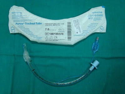
Case: A 36-year-old lady was scheduled for elective Laparoscopic Cholecystectomy. She had a history of rheumatoid arthritis with restricted neck extension. Her other airway parameters were normal. In the operating room she was preoxygenated and induced with Thiopentone Sodium and Suxamethonium . The laryngoscopic view was grade 3 and a bougie was used to facilitate intubation. An 7.0 size Portex endotracheal tube ( Fig.1)was used after routine preoperative endotracheal tube check. She was then connected to the Drager Fabius Anaesthesia machine with ventilator for the maintenance with 66 % N2O , 33 % O2 , Isofluorane and Vecuronium Bromide and the surgery started. Within few minutes of commencing positive pressure ventilation , there was the alarm for airway pressure leak , though there was no appreciable air leak from the mouth as evident by bubbling or leaking noise. The position of the tube was confirmed by equal air entry on both chest and mark at the lip level. At this stage the pilot balloon was noted to have deflated partially. The pilot balloon was inflated with 2 mls of air. After 5 minutes, the leak appeared again and the pilot balloon was again noted to have deflated. I suspected damage to the cuff and decided to replace the endotracheal tube. I then passed a bougie and railroaded a new endotracheal tube. I did not encounter any problem after that and proceeded with the surgery. On examination of the endotracheal tube, I could not find any obvious defect in the cuff or in the pilot balloon. I inflated the cuff with air, which remained patent for 5 minutes, but then started to deflate slowly. I then placed the endotracheal tube inside a water bowl and inflated the cuff (2). To my surprise, there was a leak at the junction where the inflation tube for the pilot balloon is attached to the endotracheal tube(Fig.2, 3 & 4). Even though the endotracheal tube was inspected and the cuff checked before intubation, I could not identify the leak since the cuff remained patent for few minutes. There are reports of endotracheal tube failure due to various reasons like endotracheal tube cuff failure due to valve damage (1), endotracheal tube kinking at the junction where the inflation tube for the pilot balloon is attached to the endotracheal tube (2), the problem we had was an unusual one. I would like to suggest that, during the routine checking of the endotracheal tube, the cuff should be left inflated for at least few minutes to rule out any problems like what we encountered particularly when faced with an anticipated difficult intubation.


References:
1) Heusner JE, Viscomi CM. End tracheal tube cuff failure due to valve damage. Anesth Analg. 1991; 72: 270.
2) Chua WL, Ng AS. A defective endotracheal tube, Singapore Med J. 2002; 43: 476-8.
3) Thomas A. Gettelman,Geoffrey N. Morris, Endotracheal tube failure: undetected by routine testing. Anesth Analg. 1995; 81:1313
Case Report - Endotracheal tube manufacturing defect: undetected by routine checking
Dr.Prasant Kumar Kabi, MD (Anaesthesiology)
Consultant Anaesthesiologist,
TATA MOTORS HOSPITAL,
JAMSHEDPUR, JHARKHAND, INDIA.
pkabi@tatamotors.com
Background: Routine inspection and checking of endotracheal tubes prior to use may fail to detect certain manufacturing defects. Here is a case of endotracheal tube leak at the junction where the inflation tube for the pilot balloon is attached to the endotracheal tube. This case report emphasize the importance of maintaining awareness that airway obstruction or leak due to structural defects can still occur even with high quality, prepacked single use plastic endotracheal tubes.
Case: A 36-year-old lady was scheduled for elective Laparoscopic Cholecystectomy. She had a history of rheumatoid arthritis with restricted neck extension. Her other airway parameters were normal. In the operating room she was preoxygenated and induced with Thiopentone Sodium and Suxamethonium . The laryngoscopic view was grade 3 and a bougie was used to facilitate intubation. An 7.0 size Portex endotracheal tube ( Fig.1)was used after routine preoperative endotracheal tube check. She was then connected to the Drager Fabius Anaesthesia machine with ventilator for the maintenance with 66 % N2O , 33 % O2 , Isofluorane and Vecuronium Bromide and the surgery started. Within few minutes of commencing positive pressure ventilation , there was the alarm for airway pressure leak , though there was no appreciable air leak from the mouth as evident by bubbling or leaking noise. The position of the tube was confirmed by equal air entry on both chest and mark at the lip level. At this stage the pilot balloon was noted to have deflated partially. The pilot balloon was inflated with 2 mls of air. After 5 minutes, the leak appeared again and the pilot balloon was again noted to have deflated. I suspected damage to the cuff and decided to replace the endotracheal tube. I then passed a bougie and railroaded a new endotracheal tube. I did not encounter any problem after that and proceeded with the surgery. On examination of the endotracheal tube, I could not find any obvious defect in the cuff or in the pilot balloon. I inflated the cuff with air, which remained patent for 5 minutes, but then started to deflate slowly. I then placed the endotracheal tube inside a water bowl and inflated the cuff (2). To my surprise, there was a leak at the junction where the inflation tube for the pilot balloon is attached to the endotracheal tube(Fig.2, 3 & 4). Even though the endotracheal tube was inspected and the cuff checked before intubation, I could not identify the leak since the cuff remained patent for few minutes. There are reports of endotracheal tube failure due to various reasons like endotracheal tube cuff failure due to valve damage (1), endotracheal tube kinking at the junction where the inflation tube for the pilot balloon is attached to the endotracheal tube (2), the problem we had was an unusual one. I would like to suggest that, during the routine checking of the endotracheal tube, the cuff should be left inflated for at least few minutes to rule out any problems like what we encountered particularly when faced with an anticipated difficult intubation.


References:
1) Heusner JE, Viscomi CM. End tracheal tube cuff failure due to valve damage. Anesth Analg. 1991; 72: 270.
2) Chua WL, Ng AS. A defective endotracheal tube, Singapore Med J. 2002; 43: 476-8.
3) Thomas A. Gettelman,Geoffrey N. Morris, Endotracheal tube failure: undetected by routine testing. Anesth Analg. 1995; 81:1313
Tuesday, April 8, 2008
Tuesday April 8, 2008
A brief reminder note on TRALI !!
Transfusion related acute lung injury (TRALI) is considered most important severe complication of blood transfusion probably still underdiagnosed and underreported. A lot of physicians are not aware of this and there have been several publications to inform the physicians during last 10 years or so. TRALI is defined as new acute lung injury occurring during or within 6 hrs after a transfusion, with a clear temporal relationship to the transfusion. Also, another important concept is that acute lung injury temporally associated with multiple transfusions can be TRALI, because each unit of blood or blood component can carry one or more of the possible causative agents: antileukocyte antibody, biologically active substances, and other yet unidentified agents. The treatment of TRALI is supportive, and the prognosis is generally better than for other causes of acute lung Injury. As many as one third of all patients who develop acute lung injury have been exposed to blood products. TRALI may be an important and potentially preventable cause of acute lung injury.
See excellent review Transfusion-Related Acute Lung Injury, Ognjen Gajic; S Breanndan Moore, Mayo Clinic Proceedings; Jun 2005; 80, 6; pg. 766
Previous related pearls:
TRALI
Need fresh frozen plasma but male-donor-only please !!
Reference: click to get abstract
1. Transfusion-Related Acute Lung Injury, J Intensive Care Med 2008; 23; 109
Monday, April 7, 2008
Monday April 7, 2008
Wells score of Pulmonary Embolism
Apart from Geneva and PESI, another easy score for clinical probability of Pulmonary Embolism is wells score

Click to get enlarge pic
Reference:
1. Wells PS, Hirsh J, Anderson DR, Lensing AW, Foster G, Kearon C, Weitz J, D'Ovidio R, Cogo A, Prandoni P (1995). "Accuracy of clinical assessment of deep-vein thrombosis". Lancet 345 (8961): 1326-30.
Wells score of Pulmonary Embolism
Apart from Geneva and PESI, another easy score for clinical probability of Pulmonary Embolism is wells score
Click to get enlarge pic
Reference:
1. Wells PS, Hirsh J, Anderson DR, Lensing AW, Foster G, Kearon C, Weitz J, D'Ovidio R, Cogo A, Prandoni P (1995). "Accuracy of clinical assessment of deep-vein thrombosis". Lancet 345 (8961): 1326-30.
Sunday, April 6, 2008
Sunday April 6, 2008
Pulmonary Embolism Severity Index (PESI)
Continuing our theme from yesterday on PE scores, here is another very popular index in use, call Pulmonary Embolism Severity Index (PESI). A total point score for a given patient is obtained by summing the patient's age in years and the points for each applicable characteristic.
Predictors (Points Assigned in bracket)
Demographic characteristics
Age, (one point per yr Age)
Male sex (+10)
Comorbid illnesses
Cancer (+30)
Heart failure (+10)
Chronic lung disease (+10 )
Clinical findings
Pulse more than/= 110/min (+20)
Systolic BP less than 100 mm Hg (+30)
Respiratory rate more than/=30/min (+20)
Temperature less than 36°C (+20 )
Altered mental status* (+60)
Arterial oxygen saturation less than 90% ^ (+20)
* Defined as disorientation, lethargy, stupor, or coma.
^With and without the administration of supplemental oxygen
Reference: click to get article
1. Derivation and Validation of a Prognostic Model for Pulmonary Embolism, American Journal of Respiratory and Critical Care Medicine Vol 172. pp. 1041-1046, (2005)
Pulmonary Embolism Severity Index (PESI)
Continuing our theme from yesterday on PE scores, here is another very popular index in use, call Pulmonary Embolism Severity Index (PESI). A total point score for a given patient is obtained by summing the patient's age in years and the points for each applicable characteristic.
- Score Less than/= 65 is class I (very low risk)
- Score 66–85 is class II (low risk)
- Score 86–105 is class III (intermediate risk)
- Score 106–125 is class IV (high risk)
- Score more than 125 is class V (very high risk)
Predictors (Points Assigned in bracket)
Demographic characteristics
Age, (one point per yr Age)
Male sex (+10)
Comorbid illnesses
Cancer (+30)
Heart failure (+10)
Chronic lung disease (+10 )
Clinical findings
Pulse more than/= 110/min (+20)
Systolic BP less than 100 mm Hg (+30)
Respiratory rate more than/=30/min (+20)
Temperature less than 36°C (+20 )
Altered mental status* (+60)
Arterial oxygen saturation less than 90% ^ (+20)
* Defined as disorientation, lethargy, stupor, or coma.
^With and without the administration of supplemental oxygen
Reference: click to get article
1. Derivation and Validation of a Prognostic Model for Pulmonary Embolism, American Journal of Respiratory and Critical Care Medicine Vol 172. pp. 1041-1046, (2005)
Saturday, April 5, 2008
Saturday April 5, 2008
Geneva Score for suspected pulmonary embolism (PE)
The Geneva pre-test probability score is intend to replace empirical assessment of patients with suspected pulmonary embolism (PE).
To note that literature is still raw in testing the effect of clinical experience on the inter-rater variability of Geneva as well as other scores (like PESI or Wells pre-test probability score) , and on empirical judgement, is unknown.
Total score:
- less than 5 indicates a low probability of PE
- 5–8 indicates a moderate probability of PE
- more than 8 indicates a high probability of PE
 Click to get bigger image
References: click to get article
1. Assessing clinical probability of pulmonary embolism in the emergency ward: a simple score: Arch Intern Med 2001; 161:92–97.
Friday, April 4, 2008
Friday April 4, 2008
A simple device to increase rates of compliance in maintaining 30-degree head-of-bed elevation in ventilated patients
A study published in April 2008 of 'Critical Care Medicine' to determine whether a highly visible device that clearly indicates whether the head-of-bed is adequately elevated would increase rates of compliance with head-of-bed elevation guidelines. It was a prospective, single-center, multi-unit, two-phase study of 268 intubated patients.
First phase: At the onset of the trial, nurses were reminded to maintain head-of-bed elevation more than 30 degrees. Over the subsequent 2 wks, head-of-bed elevations of intubated patient beds were measured.
Second Phase: An Angle Indicator, designed to clearly display whether the head-of-bed was adequately elevated, was then placed on side rails of beds of ventilated patients, and head-of-bed elevation measurements were taken for an additional 2 wks.
Results:
- The average head-of-bed elevation was 21.8 degrees on beds without the device, and
- 30.9 degrees on beds with the device
When compliance is defined as a bed angle of more than / = 28 degrees,
- 23% of beds without the device were compliant while
- 71.5% of the beds with the device were compliant
Nursing satisfaction
- 72% of nurses surveyed found it to be an improvement over existing methods,
- 88% found it helpful, and
- 84% would like it routinely used
Conclusions: The Angle Indicator improved rates of adherence to bed-elevation guidelines, and hospital staff found it helpful.

The Angle Indicator consists of a piece of glossy photographic printer paper cut into a pie-slice wedge. The base of the paper has a stripe of green and then red. A silk suture hangs from the apex of the wedge to its base and a steel nut is tied at the distal end of the suture. The angle indicator is placed on the side rail of the hospital bed such that when the bed is elevated to 30 degrees, the weight hangs at the junction between red and green. When the bed is elevated >30 degrees, the weight hangs in the green zone, and when the elevation is less than 30 degrees, the weight hangs in the red zone.
References:
1. A simple device to increase rates of compliance in maintaining 30-degree head-of-bed elevation in ventilated patients - Critical Care Medicine. 36(4):1155-1157, April 2008.
Thursday, April 3, 2008
Thursday April 3, 2008
Rapid Disuse Atrophy of Diaphragm Fibers in Mechanically Ventilated Humans
A very interesting study published in The New England Journal of Medicine this week.
Background: The combination of complete diaphragm inactivity and mechanical ventilation (for more than 18 hours) elicits disuse atrophy of myofibers in animals. It was hypothesized that the same may also occur in the human diaphragm.
Methods: Biopsy specimens obtained from 2 groups
Case subjects - the costal diaphragms of 14 brain-dead organ donors before organ harvest (case subjects). Case subjects had diaphragmatic inactivity and underwent mechanical ventilation for 18 to 69 hours
(compared them with)
control subjects - intraoperative biopsy specimens from the diaphragms of 8 patients who were undergoing surgery for either benign lesions or localized lung cancer (control subjects). control subjects diaphragmatic inactivity and mechanical ventilation were limited to 2 to 3 hours
Histologic, biochemical, and gene-expression studies were carried on these specimens.
Results: As compared with diaphragm-biopsy specimens from controls, specimens from case subjects showed
Conclusions: The combination of 18 to 69 hours of complete diaphragmatic inactivity and mechanical ventilation results in marked atrophy of human diaphragm myofibers. These findings are consistent with increased diaphragmatic proteolysis during inactivity.
References:
1. Rapid Disuse Atrophy of Diaphragm Fibers in Mechanically Ventilated Humans - The New England Journal of Medicine, Volume 358:1327-1335, Number 13, March 27 2008
Rapid Disuse Atrophy of Diaphragm Fibers in Mechanically Ventilated Humans
A very interesting study published in The New England Journal of Medicine this week.
Background: The combination of complete diaphragm inactivity and mechanical ventilation (for more than 18 hours) elicits disuse atrophy of myofibers in animals. It was hypothesized that the same may also occur in the human diaphragm.
Methods: Biopsy specimens obtained from 2 groups
Case subjects - the costal diaphragms of 14 brain-dead organ donors before organ harvest (case subjects). Case subjects had diaphragmatic inactivity and underwent mechanical ventilation for 18 to 69 hours
(compared them with)
control subjects - intraoperative biopsy specimens from the diaphragms of 8 patients who were undergoing surgery for either benign lesions or localized lung cancer (control subjects). control subjects diaphragmatic inactivity and mechanical ventilation were limited to 2 to 3 hours
Histologic, biochemical, and gene-expression studies were carried on these specimens.
Results: As compared with diaphragm-biopsy specimens from controls, specimens from case subjects showed
- decreased cross-sectional areas of slow-twitch and fast-twitch fibers of 57% (P=0.001) and 53% (P=0.01), respectively,
- decreased glutathione concentration of 23% (P=0.01),
- increased active caspase-3 expression of 100% (P=0.05),
- a 200% higher ratio of atrogin-1 messenger RNA (mRNA) transcripts to MBD4 (a housekeeping gene) (P=0.002), and
- a 590% higher ratio of MuRF-1 mRNA transcripts to MBD4 (P=0.001).
Conclusions: The combination of 18 to 69 hours of complete diaphragmatic inactivity and mechanical ventilation results in marked atrophy of human diaphragm myofibers. These findings are consistent with increased diaphragmatic proteolysis during inactivity.
References:
1. Rapid Disuse Atrophy of Diaphragm Fibers in Mechanically Ventilated Humans - The New England Journal of Medicine, Volume 358:1327-1335, Number 13, March 27 2008
Wednesday, April 2, 2008
Wednesday April 2, 2008
Q: You have been called by the nurse at bedside as she noticed some fresh blood in ETT. Patient has pulmonary artery catheter (PAC) in place. You are worried about pulmonary artery perforation. What would be be your first response before you initiate whole 9 yards of workup and management?
A; "Get good lung up".
First thing you need to know which pulmonary artery (Right or Left) has distal end of PAC and get patient 's position change to lateral decub. with good lung up to avoid soiling of good lung.
If patient is on ventilator, other trick is to increase PEEP (literature is controversial about its efficacy but there is no harm in increasing PEEP to control catastrophy). Overall management is supportive if pulmonary artery perforation is confirmed. Actually it is better to leave PAC intact with ballon in inflated position !! Reversal of anticoagulation if needed, hemodynamic support and preventing good lung from soiling (like applying double lumen ETT) are the initial mainstay of treatment - followed by attempt to control bleeding with interventional radiology help.
References:
1. Pulmonary Artery Rupture Induced by a Pulmonary Artery Catheter: A Case Report and Review of the Literature. University of Miami School of Medicine, Miami, Florida. Ref: Journal of Intensive Care Medicine, Vol. 19, No. 5, 291-296 (2004)
Subscribe to:
Posts (Atom)