Thursday, April 17, 2008

Thursday April 17, 2008
NIPPV for patients that fail weaning from invasive mechanical ventilation?

Very interesting study, just published today !!


The objective of this study was to evaluate the use of bi-level noninvasive positive-pressure mechanical ventilation (NPPV) for patients that fail weaning from invasive mechanical ventilation (IMV).

Methods: This experimental randomized clinical trial followed up patients undergoing IMV weaning, under ventilation for more than 48 hours, and who failed spontaneous breathing T-piece trial. Patients with contraindications to NPPV were excluded. Before T-piece placement, arterial gases, maximal inspiratory pressure, and other parameters of invasive mechanical ventilation support were measured. During the trial, respiratory rate, tidal volume, minute volume, rapid shallow breathing index, heart rate, arterial blood pressure, and peripheral oxygen saturation were measured at 1 and at 30 minutes. After failing T-piece trial, patients were randomly divided in two groups:

patients were extubated and placed on NPPV
patients returned to IMV

Results: Of 65 patients that failed T-piece trials, 28 were placed on NPPV and 37 on IMV

  • The percentage of complications in the NPPV group was lower (28.6% vs. 75.7%), with lower incidence of pneumonia and tracheotomy.
  • Length of stay in the intensive care unit and mortality were not statistically different when comparing the groups.

Conclusions: Results suggest that NPPV is a good alternative for ventilation of patients that fail initial weaning attempts. NPPV reduces the incidence of pneumonia associated with mechanical ventilation and the need for tracheotomy.

Reference: click to get article


Noninvasive mechanical ventilation may be useful in treating patients that fail weaning from invasive mechanical ventilation: a randomized clinical trial -Critical Care 2008, 12:R51

full pdf version of article is available at site

Wednesday, April 16, 2008

Wednesday April 16, 2008

Before today's pearl, here is a feedback on our pearl from April 14, 2008 regarding Stability of Norepinephrine.

"Though it is widely believed and documented that norepinephrine is less stable in saline, one recent study actually showed no difference. Please click on reference below".

Stability of norepinephrine infusions prepared in dextrose and normal saline solutions - Can J Anaesth. 2008 Mar;55(3):163-167

Surindra J. Singh, M.D.
Chief, Emergency Medicine, VAMC, Salem, VA 24153
(Dr. Singh is also one of our co-editor for this site)


Today's Pearl

Here is a quick pocket reference card for Heparin drip adjustment:


Tuesday, April 15, 2008

Tuesday April 15, 2008
Transfusion of FFP in critically ill surgical patients is associated with an increased risk of infection


We have previous studies documenting increased risk of nosocomial Infection with allogenic packed red blood cell transfusion 1. This month one retrospective study published in Critical Care Medicine shows that, also transfusion of fresh frozen plasma in critically ill surgical patients is associated with an increased risk of infection 2.
Patients: A total of 380 non-trauma patients who received fresh frozen plasma from 2004 to 2005 were compared with 2,058 nontrauma patients who did not receive fresh frozen plasma.
Results: A significant association was found between transfusion of fresh frozen plasma and

  • ventilator-associated pneumonia with shock,
  • ventilator-associated pneumonia without shock,
  • bloodstream infection with shock,
  • undifferentiated septic shock
  • The relative risk for transfusion of fresh frozen plasma and all infections was 2.99
  • The t-test revealed a significant dose-response relationship between fresh frozen plasma and infectious complications
  • Chi-square analysis showed a significant association between infection and transfusion of fresh frozen plasma in patients who did not receive concomitant red blood cell transfusion but this association was not significant in those who did receive red blood cells in addition to fresh frozen plasma.
  • The association between fresh frozen plasma and infectious complications remained significant in the multivariate model, with an odds ratio of infection per unit of fresh frozen plasma transfused equal to 1.039. This odds ratio resembled that noted for each unit of packed red blood cells, 1.074

Conclusions: Transfusion of fresh frozen plasma is associated with an increased risk of infection in critically ill patients.


References: click to get abstract

1. Impact of allogenic packed red blood cell transfusion on nosocomial infection rates in the critically ill patient - Critical Care Medicine. 30(10):2249-2254, October 2002

2. Transfusion of fresh frozen plasma in critically ill surgical patients is associated with an increased risk of infection - Critical Care Medicine. 36(4):1114-1118, April 2008.

Monday, April 14, 2008

Monday April 14, 2008
Mix for Norepinepherine


Do you know that.....NOREPINEPHRINE (LEVOPHED) is less stable in normal saline (loose its potency from oxidation). It is preferred to be mix in dextrose as the dextrose protects against oxidation of the norepinephrine and keep it active and stable.

Sunday, April 13, 2008

Sunday April 13, 2008
Quality of professional society guidelines and consensus conference statements in critical care !


See this article/study published this month in Critical Care Medicine 1, which found that " The overall quality of critical care professional society guidelines and consensus statements, as assessed by three published quality instruments, is low".
Documents focused on 1) mechanical ventilation and 2) prevention of complications of critical illness associated with mechanical ventilation were considered.


Data Extraction: Independently, two reviewers appraised the methodologic quality of each document using the Grilli, Shaneyfelt, and Appraisal of Guideline Research and Evaluation (AGREE) instruments.

Data Synthesis: Inclusion criteria were fulfilled by 13 guidelines and 12 consensus statements.
  • Adherence to current methodologic standards was low.
  • The quality of guidelines was significantly higher than consensus statements. Guidelines had higher AGREE scores compared with consensus statements.
  • Limited data suggested that guideline quality improved from 1985 to 2005.
  • Consensus statements performed poorly in the identification and interpretation of evidence and in their description of the rationale for specific recommendations.
  • Six articles reported receiving industry funding, and 15 reported on conflicts of interest (present in three articles).

Conclusions: The overall quality of critical care professional society guidelines and consensus statements, as assessed by three published quality instruments, is low. Although the quality of guidelines seems to be increasing over time, there is room for improvement, which could in turn facilitate knowledge translation and improve patient care in the intensive care unit.


Reference: click to get abstract

Quality of professional society guidelines and consensus conference statements in critical care Critical Care Medicine. 36(4):1049-1058, April 2008.

Saturday, April 12, 2008

Saturday April 12, 2008
High PEEP doesn't decrease mortality though improve oxygenation !


Role of PEEP (positive-end–expiratory pressure) in ARDS (acute respiratory distress syndrome) is well known but what level of PEEP is beneficial is still not known. Here is a very important study of 983 consecutive patients with ALI(acute lung injury) and a P/F ratio not exceeding 250 - published recently in JAMA. (85% of the 983 study patients met criteria for ARDS at enrollment).

Objective: To compare an established low-tidal-volume ventilation strategy with an experimental strategy based on the original "open-lung approach," combining low tidal volume, lung recruitment maneuvers, and high positive-end–expiratory pressure.

Interventions
The control strategy (n=508) included
  • target tidal volumes of 6 mL/kg of predicted body weight,
  • plateau airway pressures not exceeding 30 cm H2O, and
  • conventional levels of positive end-expiratory pressure

The experimental strategy (n=475) included
  • target tidal volumes of 6 mL/kg of predicted body weight, plateau pressures not exceeding 40 cm H2O,
  • recruitment maneuvers, and
  • higher positive end-expiratory pressures

Main Outcome Measure: All-cause hospital mortality

Results
  • Tidal volumes remained similar in the 2 groups,
  • Mean positive end-expiratory pressures were 14.6 cm H2O in the experimental group vs 9.8 cm H2O among controls during the first 72 hours.
  • All-cause hospital mortality rates were 36.4% and 40.4%, respectively
  • Barotrauma rates were 11.2% and 9.1%
  • The experimental group had lower rates of refractory hypoxemia (4.6% vs 10.2%), death with refractory hypoxemia (4.2% vs 8.9%), and previously defined eligible use of rescue therapies (5.1% vs 9.3%)

Conclusions: For patients with acute lung injury and acute respiratory distress syndrome, a multifaceted protocolized ventilation strategy designed to recruit and open the lung resulted in no significant difference in all-cause hospital mortality or barotrauma compared with an established low-tidal-volume protocolized ventilation strategy. This "open-lung" strategy did appear to improve secondary end points related to hypoxemia and use of rescue therapies.


Reference: click to get abstract

Ventilation Strategy Using Low Tidal Volumes, Recruitment Maneuvers, and High Positive End-Expiratory Pressure for Acute Lung Injury and Acute Respiratory Distress Syndrome, A Randomized Controlled Trial, JAMA. 2008;299(6):637-645, Feb. 2008

Friday, April 11, 2008

Friday April 11, 2008
Adulterated marijuana - source of lead poisoning


See this correspondence from Germany published this week in The New England Journal of Medicine. We are reproducing only parts but full text, tables, figures, authors and references are available free 1.

"As a consequence of strict regulations, lead intoxication has not occurred in Germany in recent decades. Recently, during a period of 3 to 4 months, 29 patients (16 to 33 years of age) were admitted to four different hospitals in the greater Leipzig area with classic signs and symptoms of lead intoxication. ......The patients presented with abdominal cramps, nausea, anemia of varying severity, and fatigue. Most patients had basophilic stippling* and a "Burton's line," ^ and some had neurologic symptoms. In other hospitals, one patient had severe encephalopathy with hallucinations and peripheral neuropathy with permanent extensor palsy in the forearm, and another patient underwent exploratory laparoscopy.

The diagnosis was quickly established, and chelation therapy was effective, but despite great efforts by health authorities and police, the source of lead could not be identified. After 8 weeks, we detected a common pattern: the patients were young, were unemployed or were students, had a history of smoking, and had body piercings. On questioning, all the patients eventually conceded that they were regular users of marijuana smoked in "joint" form or with the use of a water pipe. We recovered either half-used packages or aliquots of "home supplies" of marijuana from three patients, and we identified elemental lead by means of atomic absorptiometry and 9-tetrahydrocannabinol by means of high-pressure liquid chromatography. One package contained obvious lead particles; this strongly indicated that the lead was deliberately added to the package rather than inadvertently incorporated into the marijuana plants from contaminated soil. .....Health authorities immediately started an anonymous screening program for marijuana users. After 2 weeks, 145 persons had used this service. A total of 95 of these persons had blood lead levels that required treatment (more than 25 µg per deciliter), and some of these persons had dangerous levels of lead ( more than 80 µg per deciliter).


The current working hypothesis of the police is that because of its high specific gravity and inconspicuous grayish color, lead was used to increase the weight of street marijuana sold by the gram and thereby to maximize profits among dealers......Lead particles smoked in a joint, which can have a core temperature of 1200°C, are very effectively absorbed in the respiratory tract. The medical community, including pediatricians, should consider adulterated marijuana as a potential source of lead intoxication".


Addendum from icuroom.net:

* Basophillic Stippling
is fine, medium or coarse blue granules with uniform distribution within the red cell and represents ribosomal RNA which is precipitated during staining. It is found in conditions such as thalassemia, hemoglobinopathies, sideroblastic anemias, heavy metal poisioning and pyrimidine-5'- nucleotidase deficiency.

^ Burton's line is a blue-purplish line on the gums seen in lead poisoning.
See image of Basophillic Stippling and Burton's line below.


Reference:

Lead Poisoning Due to Adulterated Marijuana, The New England Journal of Medicine, Volume 358:1641-1642, number 15, April 10 2008

Thursday, April 10, 2008

Thursday April 10, 2008
Role of chest X-ray machine in the transmission of resistant bacteria in the ICU


See this interesting study from Drs Levin and coll. from Hadassah Hebrew University Hospital, Jerusalem, Israel and presented at 28th International Symposium on Intensive Care and Emergency Medicine, Brussels, Belgium (18–21 March 2008).

METHODS: Compliance with infection control measures was measured covertly during the performance of daily chest X-ray scans. Bacterial surface cultures were taken from the X-ray machines. An educational intervention (informing the technicians about resistant bacteria, machine culture results and correct alcohol and glove use) was instituted. Observations and machine cultures were repeated. The appearance of resistant bacteria in patient cultures was followed.

Results: Infection control practices were compared before and after the intervention.

  • Alcohol hand-rub use before patient contact increased from 12% to 25% of occasions, from 0% to 62% prior to touching the X-ray machine and from 9% to 39% before touching the next patient. Glove use also improved significantly.
  • Resistant Gram-negative bacteria grew in 12/31 (39%) preintervention X-ray machine cultures and 0/29 postintervention cultures.
  • Cultures with no bacterial growth increased from 11/31 (33%) to 22/29 (67%) pre to post intervention.
  • New occurrences of resistant Gram-negative bacteria in clinical cultures decreased from 19 in 68 patients (28%) pre intervention to 8/84 (10%) post intervention.


Conclusion: Resistant Gram-negative bacteria are found frequently on the X-ray machine, probably being transferred on technicians' hands. This represents the potential for patient-to-patient bacteria transfer. A simple infection control intervention decreases X-ray machine contamination and is associated with a decrease in the appearance of resistant bacteria in patient cultures, although causality is not proven.


Reference: click to get abstract

A potential role for the chest X-ray in the transmission of resistant bacteria in the ICU, from 28th International Symposium on Intensive Care and Emergency Medicine, Brussels, Belgium, 18-21 March 2008. Critical Care 2008, 12(Suppl 2):P10

Wednesday, April 9, 2008

Wednesday April 9, 2008
Case Report - Endotracheal tube manufacturing defect: undetected by routine checking

Dr.Prasant Kumar Kabi, MD (Anaesthesiology)
Consultant Anaesthesiologist,
TATA MOTORS HOSPITAL,
JAMSHEDPUR, JHARKHAND, INDIA.
pkabi@tatamotors.com


Background: Routine inspection and checking of endotracheal tubes prior to use may fail to detect certain manufacturing defects. Here is a case of endotracheal tube leak at the junction where the inflation tube for the pilot balloon is attached to the endotracheal tube. This case report emphasize the importance of maintaining awareness that airway obstruction or leak due to structural defects can still occur even with high quality, prepacked single use plastic endotracheal tubes.

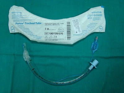
Case: A 36-year-old lady was scheduled for elective Laparoscopic Cholecystectomy. She had a history of rheumatoid arthritis with restricted neck extension. Her other airway parameters were normal. In the operating room she was preoxygenated and induced with Thiopentone Sodium and Suxamethonium . The laryngoscopic view was grade 3 and a bougie was used to facilitate intubation. An 7.0 size Portex endotracheal tube ( Fig.1)was used after routine preoperative endotracheal tube check. She was then connected to the Drager Fabius Anaesthesia machine with ventilator for the maintenance with 66 % N2O , 33 % O2 , Isofluorane and Vecuronium Bromide and the surgery started. Within few minutes of commencing positive pressure ventilation , there was the alarm for airway pressure leak , though there was no appreciable air leak from the mouth as evident by bubbling or leaking noise. The position of the tube was confirmed by equal air entry on both chest and mark at the lip level. At this stage the pilot balloon was noted to have deflated partially. The pilot balloon was inflated with 2 mls of air. After 5 minutes, the leak appeared again and the pilot balloon was again noted to have deflated. I suspected damage to the cuff and decided to replace the endotracheal tube. I then passed a bougie and railroaded a new endotracheal tube. I did not encounter any problem after that and proceeded with the surgery. On examination of the endotracheal tube, I could not find any obvious defect in the cuff or in the pilot balloon. I inflated the cuff with air, which remained patent for 5 minutes, but then started to deflate slowly. I then placed the endotracheal tube inside a water bowl and inflated the cuff (2). To my surprise, there was a leak at the junction where the inflation tube for the pilot balloon is attached to the endotracheal tube(Fig.2, 3 & 4). Even though the endotracheal tube was inspected and the cuff checked before intubation, I could not identify the leak since the cuff remained patent for few minutes. There are reports of endotracheal tube failure due to various reasons like endotracheal tube cuff failure due to valve damage (1), endotracheal tube kinking at the junction where the inflation tube for the pilot balloon is attached to the endotracheal tube (2), the problem we had was an unusual one. I would like to suggest that, during the routine checking of the endotracheal tube, the cuff should be left inflated for at least few minutes to rule out any problems like what we encountered particularly when faced with an anticipated difficult intubation.


References:

1) Heusner JE, Viscomi CM. End tracheal tube cuff failure due to valve damage. Anesth Analg. 1991; 72: 270.

2) Chua WL, Ng AS. A defective endotracheal tube, Singapore Med J. 2002; 43: 476-8.

3) Thomas A. Gettelman,Geoffrey N. Morris, Endotracheal tube failure: undetected by routine
testing. Anesth Analg. 1995; 81:1313

Tuesday, April 8, 2008

Tuesday April 8, 2008
A brief reminder note on TRALI !!

Transfusion related acute lung injury (TRALI) is considered most important severe complication of blood transfusion probably still underdiagnosed and underreported. A lot of physicians are not aware of this and there have been several publications to inform the physicians during last 10 years or so. TRALI is defined as new acute lung injury occurring during or within 6 hrs after a transfusion, with a clear temporal relationship to the transfusion. Also, another important concept is that acute lung injury temporally associated with multiple transfusions can be TRALI, because each unit of blood or blood component can carry one or more of the possible causative agents: antileukocyte antibody, biologically active substances, and other yet unidentified agents. The treatment of TRALI is supportive, and the prognosis is generally better than for other causes of acute lung Injury. As many as one third of all patients who develop acute lung injury have been exposed to blood products. TRALI may be an important and potentially preventable cause of acute lung injury.

See excellent review
Transfusion-Related Acute Lung Injury, Ognjen Gajic; S Breanndan Moore, Mayo Clinic Proceedings; Jun 2005; 80, 6; pg. 766


Previous related pearls:

TRALI

Need fresh frozen plasma but male-donor-only please !!


Reference: click to get abstract


1. Transfusion-Related Acute Lung Injury, J Intensive Care Med 2008; 23; 109

Monday, April 7, 2008

Monday April 7, 2008
Wells score of Pulmonary Embolism

Apart from Geneva and PESI, another easy score for clinical probability of Pulmonary Embolism is wells score


Click to get enlarge pic


Reference:

1. Wells PS, Hirsh J, Anderson DR, Lensing AW, Foster G, Kearon C, Weitz J, D'Ovidio R, Cogo A, Prandoni P (1995). "Accuracy of clinical assessment of deep-vein thrombosis". Lancet 345 (8961): 1326-30.

Sunday, April 6, 2008

Sunday April 6, 2008
Pulmonary Embolism Severity Index (PESI)


Continuing our theme from yesterday on PE scores, here is another very popular index in use, call Pulmonary Embolism Severity Index (PESI). A total point score for a given patient is obtained by summing the patient's age in years and the points for each applicable characteristic.

  • Score Less than/= 65 is class I (very low risk)
  • Score 66–85 is class II (low risk)
  • Score 86–105 is class III (intermediate risk)
  • Score 106–125 is class IV (high risk)
  • Score more than 125 is class V (very high risk)

Predictors (Points Assigned in bracket)

Demographic characteristics

Age, (one point per yr Age)
Male sex (+10)

Comorbid illnesses

Cancer (+30)
Heart failure (+10)
Chronic lung disease (+10 )

Clinical findings

Pulse more than/= 110/min (+20)
Systolic BP less than 100 mm Hg (+30)
Respiratory rate more than/=30/min (+20)
Temperature less than 36°C (+20 )
Altered mental status* (+60)
Arterial oxygen saturation less than 90% ^ (+20)


* Defined as disorientation, lethargy, stupor, or coma.
^With and without the administration of supplemental oxygen


Reference: click to get article

1. Derivation and Validation of a Prognostic Model for Pulmonary Embolism, American Journal of Respiratory and Critical Care Medicine Vol 172. pp. 1041-1046, (2005)

Saturday, April 5, 2008

Saturday April 5, 2008
Geneva Score for suspected pulmonary embolism (PE)

The Geneva pre-test probability score is intend to replace empirical assessment of patients with suspected pulmonary embolism (PE).

To note that literature is still raw in testing the effect of clinical experience on the inter-rater variability of Geneva as well as other scores (like PESI or Wells pre-test probability score) , and on empirical judgement, is unknown.


Total score:


  • less than 5 indicates a low probability of PE
  • 5–8 indicates a moderate probability of PE
  • more than 8 indicates a high probability of PE


Click to get bigger image

References: click to get article

1. Assessing clinical probability of pulmonary embolism in the emergency ward: a simple score: Arch Intern Med 2001; 161:92–97.

Friday, April 4, 2008

Friday April 4, 2008
A simple device to increase rates of compliance in maintaining 30-degree head-of-bed elevation in ventilated patients

A study published in April 2008 of 'Critical Care Medicine' to determine whether a highly visible device that clearly indicates whether the head-of-bed is adequately elevated would increase rates of compliance with head-of-bed elevation guidelines. It was a prospective, single-center, multi-unit, two-phase study of 268 intubated patients.


First phase: At the onset of the trial, nurses were reminded to maintain head-of-bed elevation more than 30 degrees. Over the subsequent 2 wks, head-of-bed elevations of intubated patient beds were measured.

Second Phase:
An Angle Indicator, designed to clearly display whether the head-of-bed was adequately elevated, was then placed on side rails of beds of ventilated patients, and head-of-bed elevation measurements were taken for an additional 2 wks.


Results:
  • The average head-of-bed elevation was 21.8 degrees on beds without the device, and
  • 30.9 degrees on beds with the device

When compliance is defined as a bed angle of more than / = 28 degrees,

  • 23% of beds without the device were compliant while
  • 71.5% of the beds with the device were compliant


Nursing satisfaction

  • 72% of nurses surveyed found it to be an improvement over existing methods,
  • 88% found it helpful, and
  • 84% would like it routinely used

Conclusions: The Angle Indicator improved rates of adherence to bed-elevation guidelines, and hospital staff found it helpful.


The Angle Indicator consists of a piece of glossy photographic printer paper cut into a pie-slice wedge. The base of the paper has a stripe of green and then red. A silk suture hangs from the apex of the wedge to its base and a steel nut is tied at the distal end of the suture. The angle indicator is placed on the side rail of the hospital bed such that when the bed is elevated to 30 degrees, the weight hangs at the junction between red and green. When the bed is elevated >30 degrees, the weight hangs in the green zone, and when the elevation is less than 30 degrees, the weight hangs in the red zone.


References:


1.
A simple device to increase rates of compliance in maintaining 30-degree head-of-bed elevation in ventilated patients - Critical Care Medicine. 36(4):1155-1157, April 2008.


Thursday, April 3, 2008

Thursday April 3, 2008
Rapid Disuse Atrophy of Diaphragm Fibers in Mechanically Ventilated Humans

A very interesting study published in The New England Journal of Medicine this week.


Background: The combination of complete diaphragm inactivity and mechanical ventilation (for more than 18 hours) elicits disuse atrophy of myofibers in animals. It was hypothesized that the same may also occur in the human diaphragm.

Methods: Biopsy specimens obtained from 2 groups

Case subjects - the costal diaphragms of 14 brain-dead organ donors before organ harvest (case subjects). Case subjects had diaphragmatic inactivity and underwent mechanical ventilation for 18 to 69 hours

(compared them with)


control subjects - intraoperative biopsy specimens from the diaphragms of 8 patients who were undergoing surgery for either benign lesions or localized lung cancer (control subjects). control subjects diaphragmatic inactivity and mechanical ventilation were limited to 2 to 3 hours

Histologic, biochemical, and gene-expression studies were carried on these specimens.

Results: As compared with diaphragm-biopsy specimens from controls, specimens from case subjects showed
  • decreased cross-sectional areas of slow-twitch and fast-twitch fibers of 57% (P=0.001) and 53% (P=0.01), respectively,
  • decreased glutathione concentration of 23% (P=0.01),
  • increased active caspase-3 expression of 100% (P=0.05),
  • a 200% higher ratio of atrogin-1 messenger RNA (mRNA) transcripts to MBD4 (a housekeeping gene) (P=0.002), and
  • a 590% higher ratio of MuRF-1 mRNA transcripts to MBD4 (P=0.001).

Conclusions: The combination of 18 to 69 hours of complete diaphragmatic inactivity and mechanical ventilation results in marked atrophy of human diaphragm myofibers. These findings are consistent with increased diaphragmatic proteolysis during inactivity.


References:

1. Rapid Disuse Atrophy of Diaphragm Fibers in Mechanically Ventilated Humans - The New England Journal of Medicine, Volume 358:1327-1335, Number 13, March 27 2008

Wednesday, April 2, 2008

Wednesday April 2, 2008


Q: You have been called by the nurse at bedside as she noticed some fresh blood in ETT. Patient has pulmonary artery catheter (PAC) in place. You are worried about pulmonary artery perforation. What would be be your first response before you initiate whole 9 yards of workup and management?


A; "Get good lung up".

First thing you need to know which pulmonary artery (Right or Left) has distal end of PAC and get patient 's position change to lateral decub. with good lung up to avoid soiling of good lung.

If patient is on ventilator, other trick is to increase PEEP (literature is controversial about its efficacy but there is no harm in increasing PEEP to control catastrophy). Overall management is supportive if pulmonary artery perforation is confirmed. Actually it is better to leave PAC intact with ballon in inflated position !! Reversal of anticoagulation if needed, hemodynamic support and preventing good lung from soiling (like applying double lumen ETT) are the initial mainstay of treatment - followed by attempt to control bleeding with interventional radiology help.


References:

1.
Pulmonary Artery Rupture Induced by a Pulmonary Artery Catheter: A Case Report and Review of the Literature. University of Miami School of Medicine, Miami, Florida. Ref: Journal of Intensive Care Medicine, Vol. 19, No. 5, 291-296 (2004)